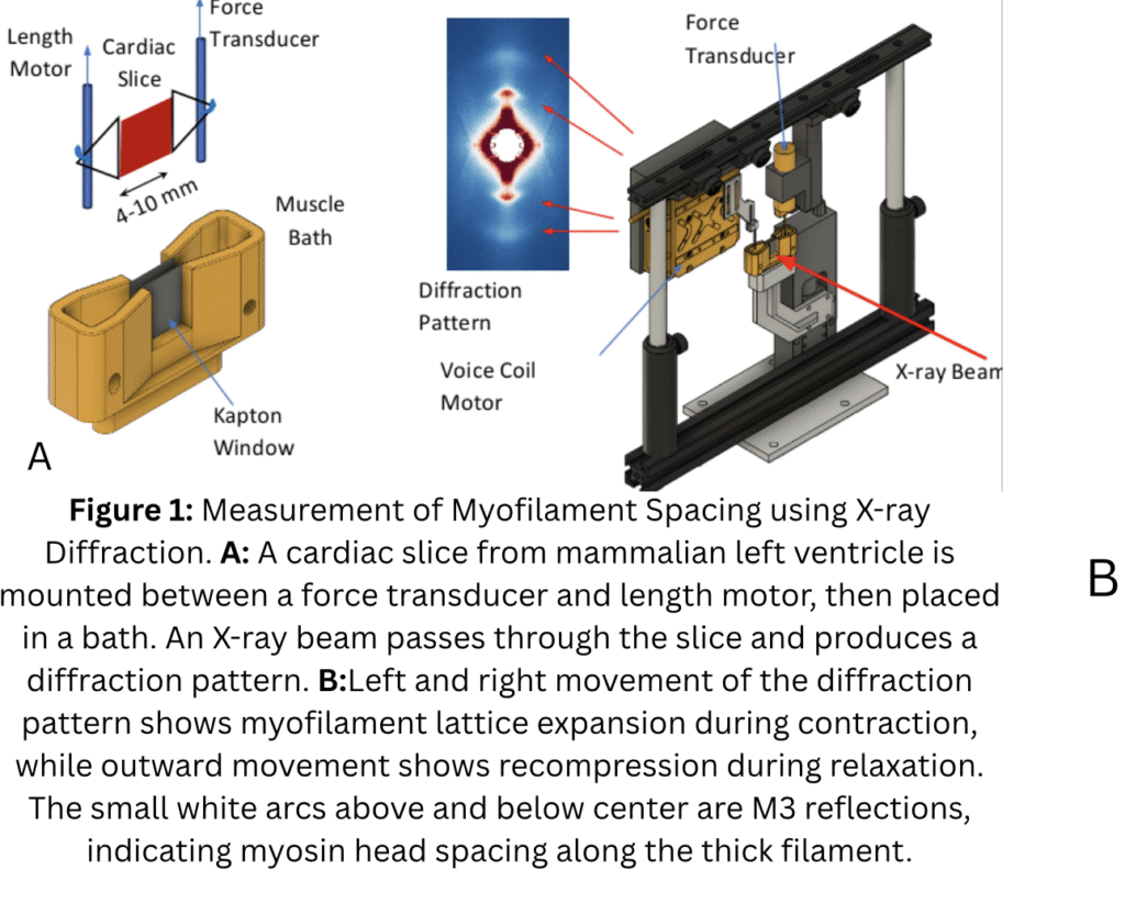
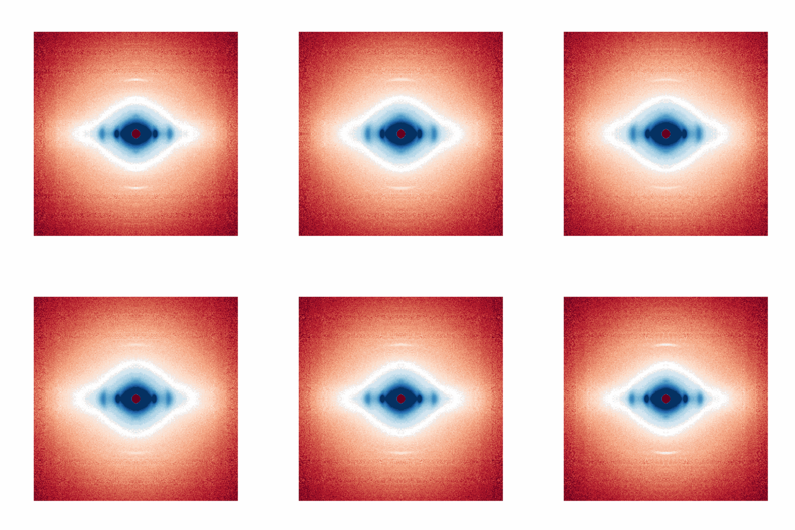
Measurement of Myofilament Spacing using X-ray Diffraction: Cardiac slices (300 µm thick) are prepared from mammalian left ventricle and mounted between a force transducer and length motor. The slice is placed in a bath and electrically stimulated while an X-ray beam passes through, producing a diffraction pattern. This pattern allows us to determine the lattice spacing of the myofilaments—the distance between thick and thin filaments—which changes during contraction and relaxation, as well as in disease states. X-ray video crystallography simultaneously detects both functional and structural dynamics in cardiac tissue, providing a unique tool for evaluating the effects of therapies.